 Do you enjoy helping others? Want to work with patients but don’t want to perform clinical tasks? As a medical sonographer, you won’t perform phlebotomy. Still, you will prepare patients to take images of their bodies with an ultrasound. The best part is that you will assist doctors in diagnosing disorders and providing the medical care your patients need.
What is a Medical Ultrasound?
An ultrasound machine is a non-invasive diagnostic equipment that uses sound waves to produce images of structures and organs within the body. Ultrasound probes produce sound waves with high frequencies beyond humans’ hearing but are visible with ultrasound equipment.
Ultrasound waves are produced by a transducer element when an electrical field is applied. These waves enter the body and are reflected from the surfaces of the organs to the transducers, where the sound waves returning to the element are changed to electrical signals that the ultrasound system will analyze and produce an image. The ultrasound system will generate a two-dimensional image of the tissues and organs.
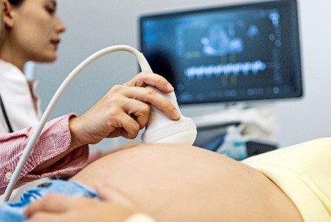
What Does a Diagnostic Medical Sonographer Do?
Diagnostic medical sonographers operate ultrasound equipment to create images of the body to help doctors and surgeons diagnose and treat diseases and disorders. Their duties include:
Preparing the Exam Room
The diagnostic medical sonographer cleans and disinfects the exam room, including the ultrasound machine, exam chair, and surfaces within the room. This process gets repeated between patients for each patient’s safety.
Managing Patients
The diagnostic medical sonographer meets and greets the patient in the waiting room. They walk the patient back to the ultrasound room. The sonographer educates the patient about what they will experience during the procedure and any associated risks. They properly drape and position the patient to get the best images. At the end of the process, the sonographer cleans off any ultrasound gel, allowing the patient to get situated before leaving the facility or speaking with the doctor.
Reviewing Images for Abnormalities
The diagnostic medical sonographer will review the images, note any abnormalities, and check for the quality of the area imaged. They cannot diagnose the patient based on the image but relay technical impressions to the radiologist.
Findings and Diagnosis
The images are prepared for the radiologist to provide a final impression. All imaging findings are recorded in the patient’s electronic health records. Then, it is up to the doctor or surgeon to review the ultrasound images to make diagnoses and treatment plans for the patient.
How Do You Become a Diagnostic Medical Sonographer?
There are a few steps to becoming a registered diagnostic medical sonographer. These steps include:
Step #1: Finding the Diagnostic Medical Sonographer Program That is Right for You
Step #2: Complete the Program
Step #3: Apply for Registration from the American Registry for Diagnostic Medical Sonography (ARDMS) or American Registry for Radiologic Technology (ARRT) credential
Step #4: Let FNU Help You Find a Job in South Florida
What is the ARDMS and ARRT?
ARDMS and ARRT are two independent credentialing organizations that administer registry examinations in ultrasound. Registered with one of these entities provides additional validation about a sonographer’s capabilities, which can help get a job.
What Do You Learn During a Medical Sonographer Program?
In addition to general education courses in Math and English, students of the Diagnostic Medical Sonography program also study:
Anatomy and Physiology
This course focuses on the 11 body systems that work together to protect the body, offer support and stability, move the body, circulate blood, and process food, for example. The 11 body systems include:
- Respiratory – helps the body breathe through the lungs and trachea.
- Cardiovascular – circulates blood within the heart, arteries, and veins
- Digestive – processes food in the mouth, stomach, intestines, and other digestive organs
- Endocrine – produces hormones using the thyroid, pituitary, and adrenal glands
- Urinary – eliminates waste from the body in the kidneys and bladder
- Nervous – coordinates all body systems through communication in the brain and nerves.
- Reproductive – helps reproduction within the ovaries and fallopian tubes
- Integumentary – protects the body from damage using skin, hair, and nails
- Muscular/Skeletal – provides form, support, stability, and movement using the muscles and bones of the body
- Lymphatic – produces blood, maintains fluid balance, and defends against disease.
Medical Terminology
This course focuses on medical terminology specific to basic human systems that you will use as a sonographer. By reviewing roots, suffixes, and prefixes, you can better understand the individual parts of a word, which will help you memorize medical terminology.
Intro to Sonography and Patient Care
A review of the fundamental sonography knowledge, this course covers the skills required to perform imaging modalities in the clinical setting with proper patient care in mind.
Cross Sectional Anatomy
This course covers the sectional anatomy of the body in the transverse, longitudinal, and coronal planes. This course will focus on the body parts imaged by sonography.
Intro to Sonographic Physics
A review of the fundamental principles of physics needed to understand clinical ultrasound, including:
- Frequency – the number of waves that pass through the body in a unit of time
- Propagation Speed – the distance the wave travels within a given time. One wavelength at a time of one period.
- Pulsed ultrasound – the waves travel into tissue and reflect to the probe at a rate determined by the tissue’s consistency.
- Wave Interaction – how waves interact with the body, including reflection, refraction, diffraction, and interference.
- Angle of Incidence – the ultrasound beam angle and the tissue plane.
- Attenuation – the reduction of intensity of a sonography wave as it moves through the body.
Abdominal Sonography
This course focuses on the abdominal anatomy, including the liver, gallbladder, pancreas, bile ducts, spleen, and abdominal aorta.
Obstetrics/Gynecology Sonography
A course in how to identify and examine the female reproductive system, monitor a fetus, and apply obstetrical ultrasound.
Small Parts
This course reviews the breast and the basics of the thyroid, parathyroid, neck glands, scrotum, and prostate, including basic anatomy, scanning techniques, and expectations.
Neonatal and Pediatric Sonography
A course on the imaging fundamentals and scanning techniques of the adolescent and infant abdominal neurosonography, neonatal spine, and pediatric hip.
Intro to Echocardiography (ECG)
An introduction to the procedures and techniques of imaging cardiac anatomy in a non-invasive method. The ECG is used to offer a thin cross-section of the cardiac structure.
Intro to Vascular Technology
An introduction to the vascular system and the use of an ultrasound that produces images of the veins and arteries using high-frequency sound waves.
Eager to Learn More?
The Diagnostic Medical Sonographer Technology Program will provide you with the knowledge and skills necessary to acquire the technical competencies to function as an entry-level Diagnostic Medical Sonographer. The curriculum that is followed in this Program complies with the National Educational Curriculum for Sonography (NEC) outline and accreditation agencies to provide competency-based, outcome-oriented, didactic, laboratory, and clinical experience as it relates to the general concentration, which includes Abdomen, obstetrics, and gynecology (OB/GYN).
Let Florida National University answer any questions if you are interested in a Diagnostic Medical Sonography career. Contact us today to learn more about our Diagnostic Medical Sonographer Technology Degree programs.